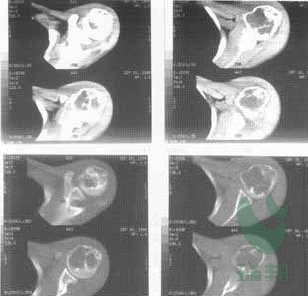
病例三 左肱骨近端巨细胞瘤
 |
|
| fig1 | |
 |
 |
| fig2 | fig3 |
 |
 |
| fig4 | fig5 |
 |
|
| fig6 | |
1. Before operation, CT scanning
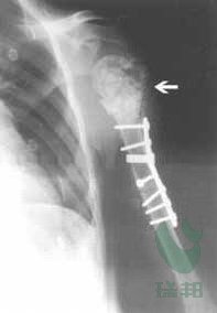
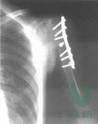
2. The defect was filled with CPC granules after the clearance of focal lesion
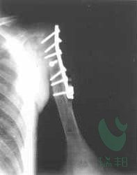
3. 3 months later, the density of the filling position began lower, which indicated the material began to degrade gradually
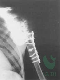
4. 6 months later
5. 9 months later
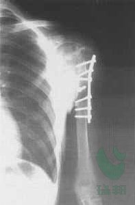
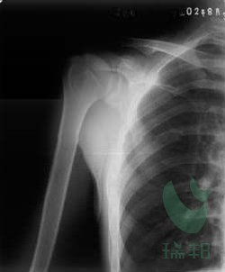
6. 12 months later
 |
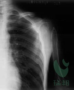 |
| After cured,the comparion between the left humerus and the right humerus | |